+++ Meet us in person
+++
26th Federal Congress Pathology, Berlin, October 2–3, 2026
+++ Meet us in person +++ 26th Federal Congress Pathology, Berlin, October 2–3, 2026
Native Digital Histology:
THE FUTURE OF
TISSUE DIAGNOSTICS
The world’s first tissue-preserving, fully automated digital histopathology solution for AI-assisted diagnostics.


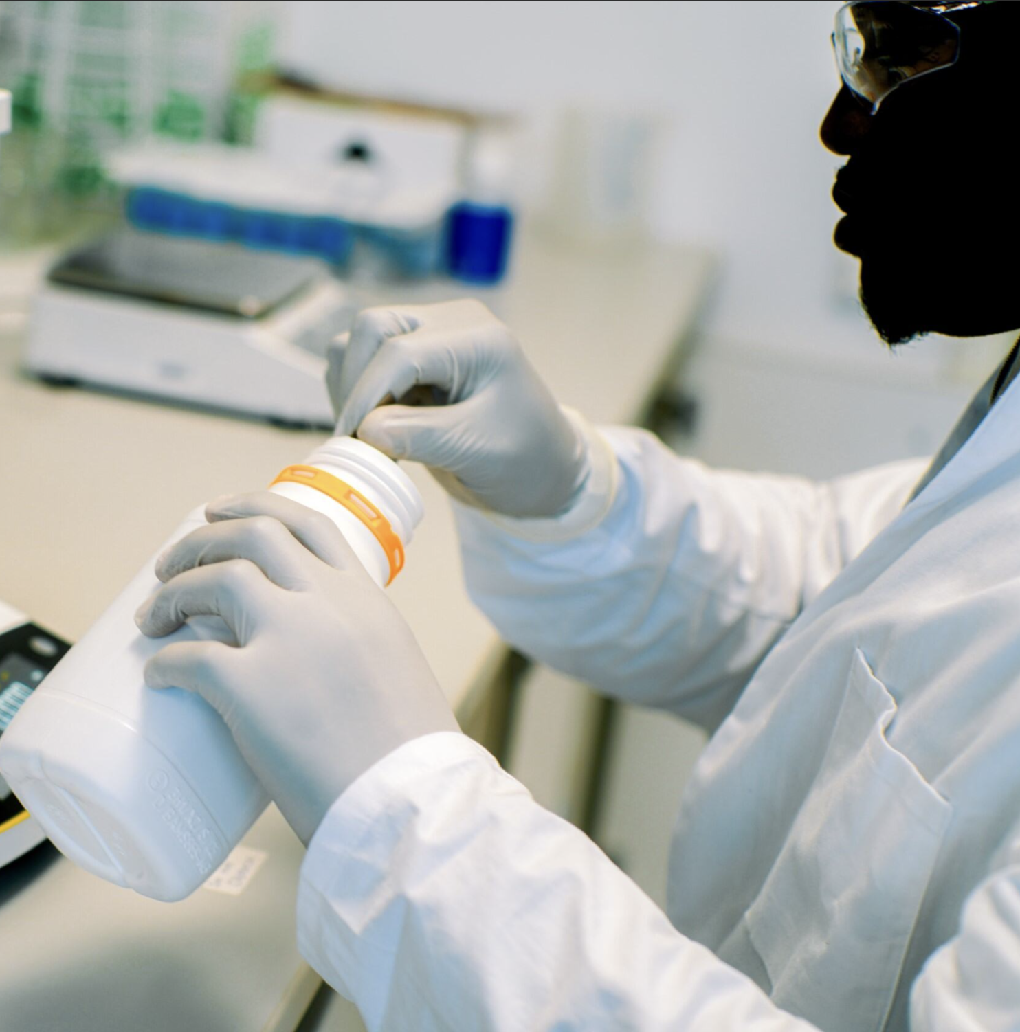
The Challenge in Tissue Diagnostics
Histopathology is a cornerstone of modern medicine. Simultaneously, demands for depth of data, process consistency, swift turnaround times, and the integration of morphological and molecular information are rising.
Standard histopathology methods often exhaust precious tissue samples, hindering comprehensive genomic and proteomic profiling. We are bridging the gap in the diagnostic process and unlocking the potential of precision oncology.
-
Precision medicine increasingly requires a holistic view of tissue architecture, spatial information, and molecular analysis.
-
High caseloads, numerous process steps, and limited personnel resources define everyday life in diagnostic labs.
-
Future diagnostic processes require data that can be consistently reused and integrated digitally.
Our Solution:
Native Digital Histology
From tissue sample to diagnosis – fully automated.
LIMAA combines proprietary reagents, automated processing, and software into an integrated solution. This creates a consistent journey from the sample to native digital tissue imaging and advanced analysis.
Proprietary reagents for optimal tissue staining and preparation. Specially developed for native digital imaging with the highest color consistency and reproducibility.
Reagents
Fully automated system for tissue processing. No manual cutting, no scanning of glass slides – direct digital capture in an integrated workflow.
Hardware

AI-powered image analysis and seamless integration into existing pathology information systems. Automated diagnostic support for faster, more accurate diagnoses.
Software & AI
We do not digitalize the current workflow.
We replace it.
Digitized pathology
Manual workflow with over 15 individual steps – time-consuming and labor-intensive.
PROCESS
Glass slides must be scanned – additional sources of error, and additional costs.
IMAGE TYPE
CONSISTENCY
High variability: Hardware and Inter-observer
TISSUE
Limited, due to consumption
LIMAA Solution
PROCESS
Fully automated process – from sample to diagnosis in one system
IMAGE TYPE
Native digital tissue images for highest image quality
Standardized and AI-ready
CONSISTENCY
Preserved, ready for multi-omics
TISSUE
Why Tissue-Preserving Microscopy is Key
-
AI-supported evaluation and workflow automation link morphological, spatial and molecular data into an integrated analysis process.
-
Volumetric microscopy combines classical histopathology with modern imaging and improves laboratory processes.
-
More precise diagnoses and treatment decisions through the integration of tissue and molecular diagnostics – the basis for personalized medicine.
Advanced Diagnostics:
Our Nanobody Portfolio.
To further expand the diagnostic potential of our histology workflow, we offer a dedicated library of proprietary nanobodies. Designed for high-specificity labeling, these tools unlock new dimensions for multiplexing in pathology.
NEWS

What People Say About Us.
-
![Prof. Dr. Bruno Maerkl, smiling in white lab coat, blue shirt and tie, professional portrait in medical setting]()
“As a pathologist, I have always struggled with the limitations of traditional histopathology methods, especially the manual processes that often result in tissue loss and longer turnaround times. Integrating LIMAA's fully automated, tissue-preserving histopathology solution into our workflow will transform it remarkably. Overall, LIMAA's innovative technology is not just a step forward; it's a leap into the future of histopathology.”
Prof. Dr. Bruno Märkl
Director of the Institute of Pathology and Molecular Diagnostics, University Hospital Augsburg -
![Prof. Dr. med. Jörg Debatin, new Chairman of LIMAA Technologies Board, smiling professional portrait in light blue shirt against office background]()
“I am looking forward to working with a top-notch team of scientists and engineers dedicated to fundamentally improving efficiency and accuracy of tissue-based diagnosis.’’
Prof. Dr. med. Jörg Debatin, MBA
Healthcare Entrepreneur -
![René Hägerling, smiling professional portrait on gray background, wearing light blue shirt, LIMAA Technologies team member.]()
“Thanks to our sample preparation methods and advanced light-sheet microscopy, we can visualize entire tissue samples in 3D without physically sectioning them. Our ability to perform rapid optical sectioning and obtain thousands of digital images in seconds or minutes has significantly increased our throughput.”
Dr. Dr. René Hägerling
Co-Founder of LIMAA Technologies


“LIMAA was founded at Charité – one of the world’s leading medical institutions – because a group of scientists refused to accept that a 150-year-old workflow was sufficient for 21st-century medicine.”
Join us in shaping the Future of Tissue Diagnostics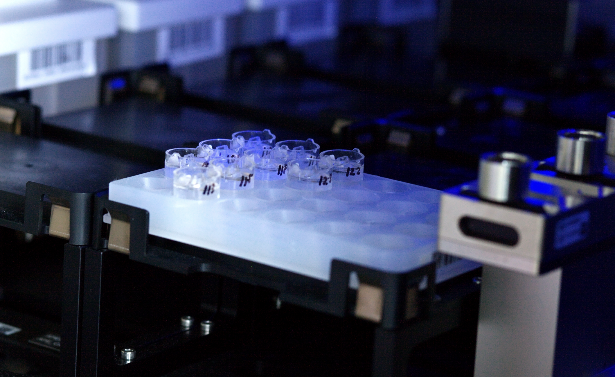
MORE
INSIGHTS IN
LESS TIME.