
Publication: 3D Visualization of Human Blood Vascular Networks
We just published one essential part of our core technology – single-domain antibodies for 3D visualization of biomarkers:
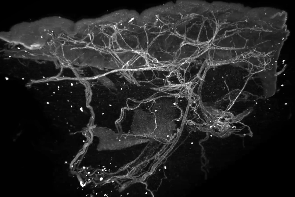
High-quality three-dimensional (3D) microscopy allows detailed, unrestricted and non-destructive imaging of entire volumetric tissue specimens and can therefore increase the diagnostic accuracy of histopathological tissue analysis. However, commonly used IgG antibodies are oftentimes not applicable to 3D imaging, due to their relatively large size and consequently inadequate tissue penetration and penetration speed. […]
Here, we report the generation and experimental validation of single-domain antibodies directed against the human endothelial cell-selective adhesion molecule (hESAM), which enables spatial visualization of blood vascular networks in whole-mount 3D imaging. After analysis of Nb binding properties and quality, selected Nb clones were validated in 2D and 3D imaging approaches, demonstrating comparable staining qualities to commercially available hESAM antibodies in 2D, as well as rapid and complete staining of entire specimens in 3D.
Please access the entire publication via the link below:
International Journal of Molecular Sciences, March 2022
Share this post: