
Meet us in person
+++
109th Annual Meeting of DGP, Augsburg, May 28 - 30, 2026
+++
26th Federal Congress Pathology Berlin, October 2 - 3, 2026
Meet us in person +++ 109th Annual Meeting of DGP, Augsburg, May 28 - 30, 2026 +++ 26th Federal Congress Pathology Berlin, October 2 - 3, 2026
World’s First Fully Automated Tissue Preserving Histopathology Platform
The Platform Enables the Complete Automation of the Pathology Sample Process.
What we doAt LIMAA Technologies, we develop a fully automated and scalable tissue preserving end-to-end 3D histopathology platform for your clinical use. The platform allows you to analyze entire tissue specimens without embedding or slicing.
OUR INNOVATION
LIMAA Speeds Up Turnaround Time by
Directly Processing and Digitalizing Entire Tissue Samples
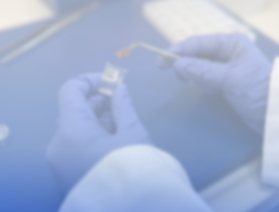

Sample
Preparation
STEP 1
Based on 13+ years experience in reagent development for light-sheet microscopy, we develop standardized fluorescent histochemical- and immuno-stainings for a swift pre-analytical process. Tissue samples remain intact and can be used for genomics, in each and every case.
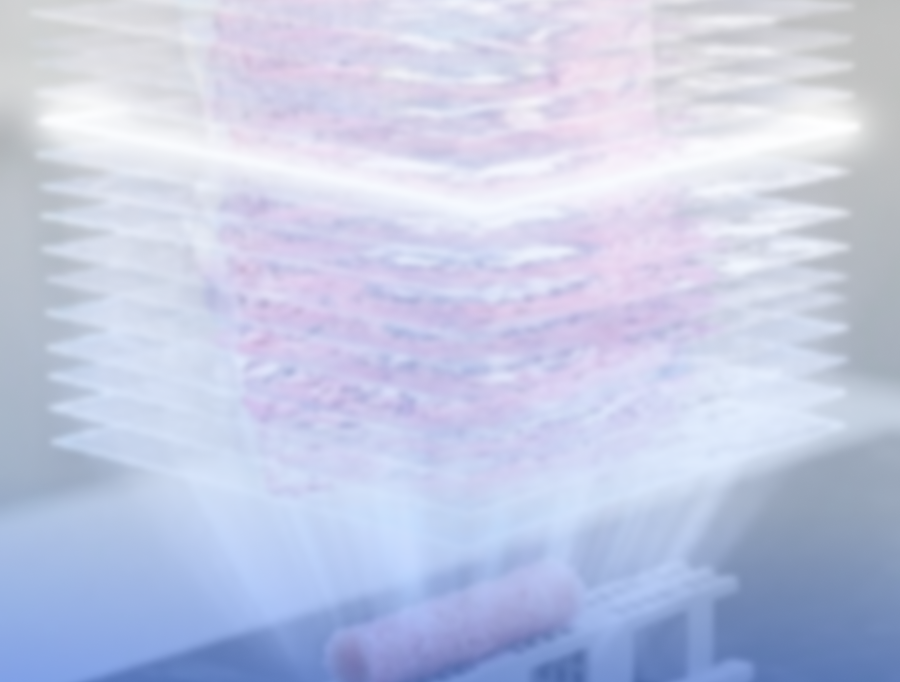
Automation
and Digitalization
STEP 2
At LIMAA Technologies, we fully automate Sample Preparation and Digitalization using Light-Sheet Microscopy reducing Turn-Around-Time by 50% and hardware costs for digitization by 65%.
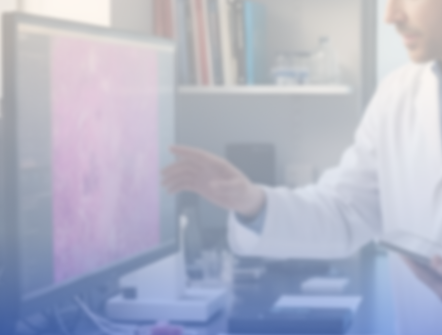
Digital
Diagnosis
STEP 3
Since digital pictures come for free in our process, we directly augment analysis and quality control of samples in the background, enabling you as a pathologist to do your most important job - diagnosis and guiding therapy options.
News


PRODUCT
Key Features
Fully
Automated

Replicable & High-Throughput
Avoid Human-Error
Digital Workflow and Diagnosis

High Sensitivity and Specificity
Seamless Integration
Tissue Preserving Analysis

Cover Entire Tissue Specimens
Integration of NGS in Each Sample
Prepared for
Future Challenges

Flexible Integration of Assays
Scalable Process
What People Say About Us.
-
![Prof. Dr. Bruno Maerkl, smiling in white lab coat, blue shirt and tie, professional portrait in medical setting]()
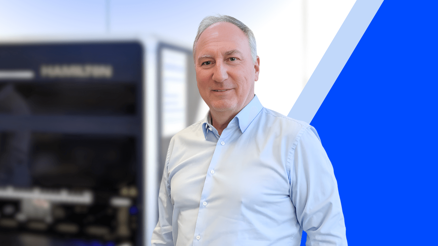
“As a pathologist, I have always struggled with the limitations of traditional histopathology methods, especially the manual processes that often result in tissue loss and longer turnaround times. Integrating LIMAA's fully automated, tissue-preserving histopathology solution into our workflow will transform it remarkably. Overall, LIMAA's innovative technology is not just a step forward; it's a leap into the future of histopathology.”
Prof. Dr. Bruno Märkl
Director of the Institute of Pathology and Molecular Diagnostics, University Hospital Augsburg -
![Prof. Dr. med. Jörg Debatin, new Chairman of LIMAA Technologies Board, smiling professional portrait in light blue shirt against office background]()
“I am looking forward to working with a top-notch team of scientists and engineers dedicated to fundamentally improving efficiency and accuracy of tissue-based diagnosis.’’
Prof. Dr. med. Jörg Debatin, MBA
Healthcare Entrepreneur -
![René Hägerling, smiling professional portrait on gray background, wearing light blue shirt, LIMAA Technologies team member.]()
“Thanks to our sample preparation methods and advanced light-sheet microscopy, we can visualize entire tissue samples in 3D without physically sectioning them. Our ability to perform rapid optical sectioning and obtain thousands of digital images in seconds or minutes has significantly increased our throughput.”
Dr. Dr. René Hägerling
Co-Founder of LIMAA Technologies


32
Seconds for Visualization
Tissue conserving visualization of entire blocks within seconds based on light-sheet microscope technology and the ability to fully reuse tissue for further biomarker staining or genomics.
100%
Percent Integration
Combination of Visualization and Genomics with full integration into Pathology Workflow and Laboratory Information Management Systems including Track & Trace of sample preparation process, analysis stage, and archiving.
185
Minutes Sample Preparation
Hands-off swift and fully-automated sample preparation for routine lab stainings of entire tissue blocks without the requirement of slicing and transfer of tissue onto glass slides.
25
Computer Vision Algorithms
Direct acquisition of digital pictures and 3D volumes, quality control and guided analysis using state of the art data science and machine learning algorithms to increase accuracy and speed of diagnosis.
FAQs
What services do you offer?
1
We offer a range of solutions designed to meet your needs—whether you're just getting started or scaling something bigger. Everything is tailored to help you move forward with clarity and confidence.
How do I get started?
2
Getting started is simple. Reach out through our contact form or schedule a call—we’ll walk you through the next steps and answer any questions along the way.
What makes you different?
3
We combine a thoughtful, human-centered approach with clear communication and reliable results. It’s not just what we do—it’s how we do it that sets us apart.
How can I contact you?
4
You can reach us anytime via our contact page or email. We aim to respond quickly—usually within one business day.